MRI Datasets for Direct Download
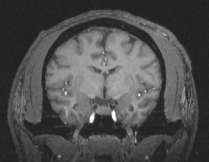
Representative Sample of Adult Chimpanzee 3.0T MRI Scans (5 males, 5 females) - Chimpanzees were MRI scanned during their annual physical examinations following standard procedures designed to minimize stress. The animals were first sedated with ketamine (10 mg/kg) or telazol (3-5mg/kg) and were subsequently anaesthetized with propofol (40–60 mg/(kg/h)). They were then transported to the MRI scanning facility and placed in a supine position in a 3.0T scanner (Siemens Trio, Siemens Medical Solutions USA, Inc., Malvern, Pennsylvania) with their head in a human-head coil. If use of these data results in publication, please give reference to the National Chimpanzee Brain Resource (supported by NIH NS092988) in the methods section and the acknowledgements of manuscript. After publication, deposit any derived data to the NCBR Data Repository.

chimpanzee.
Comparative Sample of Anthropoid Primate 1.5T MRI Scans - Whole brain MRI scans were collected from 38 living, nonhuman anthropoid primates spanning 10 species. All scans were acquired at 1.5T. Prior to scanning, nonhuman primates were anesthetized with Ketamine (10 mg/kg) and then weighed. Monkeys were scanned in a prone position with a human knee coil, whereas apes were scanned in a supine position using a human head coil. Throughout the scan, nonhuman primates received a continuous IV infusion of propofol (10–20 mg/kg/hr) for anesthesia. Head motion was minimized by stabilizing with foam cushions and elastic straps. T1-weighted images of the entire brain were acquired with a 1·5 Tesla Phillips NT scanner (Philips Medical Systems, The Netherlands) using a gradient-echo protocol with the following parameters: slice thickness=1·2 mm, slice interval=0·6 mm (in most cases), TR=19·0 msec, TE=8·5 msec, number of signals averaged=8, matrix=256_256 pixels. Field of view (FOV) was adjusted to include the entire brain in each subject, and so varied as a function of brain size. Consequently, pixel size ranged from 0·47 mm2 to 0·78 mm2. Scan duration was a function of brain size. Approximate scanning time for monkeys and apes were 40 min and 60 min, respectively. An additional inversion recovery scan was also acquired from a sub-set of animals (see scan parameters in chart). These scans were provided to the NCBR for public access courtesy of Dr. James Rilling.
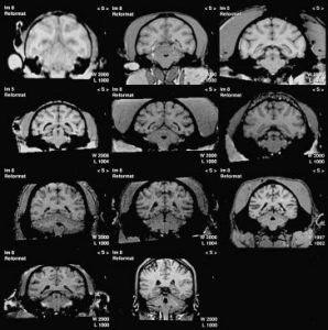
squirrel monkey, capuchin monkey, rhesus
macaque, sootey mangabey, baboon, gibbon,
orangutan, gorilla, chimpanzee, bonobo, human
(Rilling and Insel, 1999).
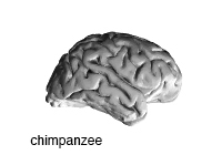
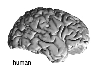
STL Files of Chimpanzee and Human Brains to Print 3D Models - Click on the images of the brains to download.