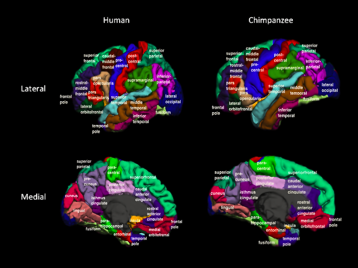
Cortical parcellation of the chimpanzee brain compared to human. Created in FreeSurfer software using a modification of the human Desikan-Killiany atlas.
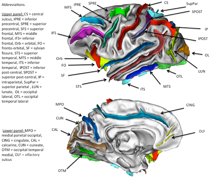
Cerebral sulci in the chimpanzee brain as segmented by BrainVisa software.
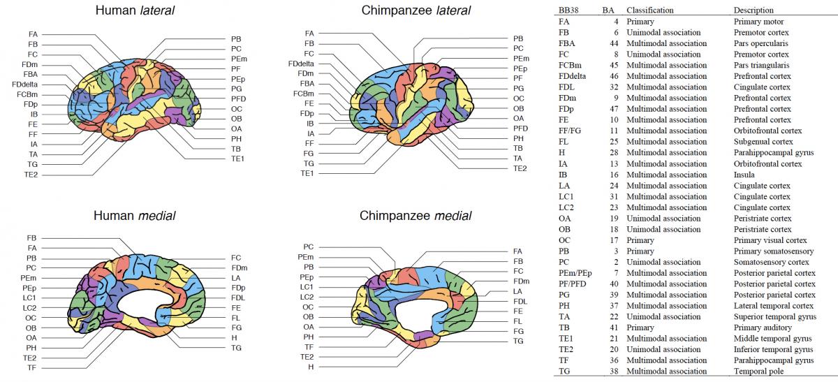
Comparative cortical maps of humans and chimpanzees, classified into primary, unimodal association, and multimodal association areas, from Ardesch et al. (2019). First column describes the cortical areas according to Bailey P, von Bonin G, McCulloch WS (1950) The isocortex of the chimpanzee (University of Illinois Press, Urbana). For reference, corresponding Brodmann areas (BA) are listed in the second column. The third column describes the classification into primary, unimodal association, and multimodal association areas according to Mesulam M-M (1998) From sensation to cognition. Brain 121:1013–1052. The Mesulam division consists of five categories: 1) primary unimodal, 2) unimodal association, 3) heteromodal/multimodal association, 4) paralimbic, and 5) limbic areas. With both heteromodal/multimodal and paralimbic areas described as processing multimodal information, categories 3 and 4 were collapsed into the class of multimodal association areas. Limbic areas were not part of the Bailey et al. (1950) cortical parcellation.
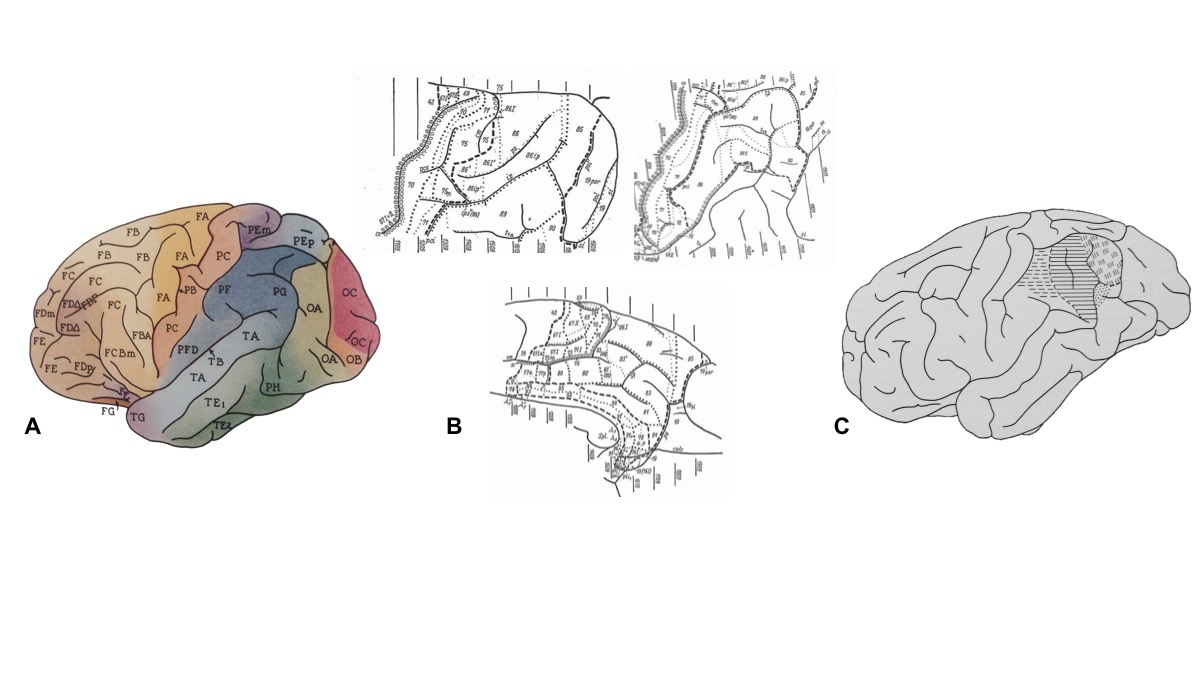
Parcellations of the chimpanzee brain. Bailey and von Bonin (1950, A) is the most widely used parcellation of the chimpanzee brain. Other parcellations include the cortical maps that were produced of the parietal lobe by Gerhardt (1938, B) and the inferior parietal lobe by Shevchenko (1936, C).